SXM II end-station
The Scanning X-ray Microscope was refurbished in 2008 to offer enhanced functionalitites, which are described hereafter.
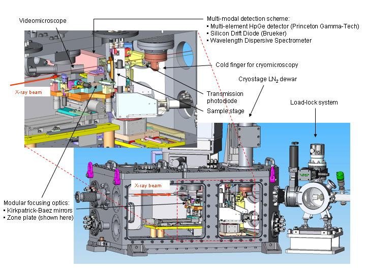
3D view of the ID21 Scanning X-ray microscope
Microscope chamber
The X-ray microscope is housed in an environmental chamber allowing operation in air or vacuum (10-4-10-6 mbar). A Kapton exit window separates the microscope environment from the beamline vacuum. A load-lock system allows fast exchange of samples under vacuum, and greatly facilitates operation under cryogenic conditions.
Focusing optics
The microscope can now host two different optical focusing configurations: either zone plates (ZP) or a Kirkpatrick-Baez mirror system (KB), which routinely achieves a typical spot size of 0.35 x 0.7 µm2 with a photon flux of 109-1010 photons/s. The KB system has been developed at ESRF and is based on elliptically shaped fixed-focus Ni coated mirrors. Its compact design ensures high mechanical / thermal stability and a short focal length for high source demagnification. The advantages of the KB configuration are its higher photon flux and its achromaticity which is preferable for µXANES
Detectors
The SXM can be equipped with a single or a 7-element HpGe fluorescence detector (Princeton Gamma-Tech, US), which offers an increased solid angle for an optimized collection of fluorescence photons, and a large area (80 mm2) XFlash 5100 Bruker Silicon Drift Diode (SDD). A compact x-ray wavelength dispersive spectrometer achieving a few tens eV energy resolution has also been implemented for highly selective fluorescence detection.
Sample stage and sample environments
The sample is scanned using a combination of piezo driven flexure and mechanical stages giving a total scan area of 20 x 20 mm2.
The sample support comprises two translation stages. A system of coarse mechanical xy-stages driven by stepper motors allow sample alignment and scanning over a range of +/-10 mm with measured resolutions of 1µm. For finer scanning resolutions a commercial piezo-driven monolithic xy-flexure stage (Physik Instrumente) allows sample scanning over a range of 100 µm. This system, equipped with integrated capacitive encoders allows positioning resolutions of about 10nm.
The sample stage can accommodate various sample holders depending on the nature of the sample (solid, liquid or frozen). Please see the sample preparation and cryo-stage sections for further details.
Sample visualisation and control of the microscope
A visible light video-microscope allows visualisation of the sample, even under vacuum, for precise alignment in the beam.
Control of the SXM is now performed through a fully integrated graphical user interface "Daquiri" which includes on-line visualization of the sample through a video-microscope, and direct “grabbing” of regions to be scanned from a video image.

partners


European Synchrotron Radiation Facility - 71, avenue des Martyrs, CS 40220, 38043 Grenoble Cedex 9, France.