- Home
- News
- General News
- A tough egg to crack...
A tough egg to crack - oldest lizard embryos discovered in fossil eggs
10-07-2015
Since their discovery in Thailand in 2003, scientists have puzzled over the identity of a batch of tiny fossilised eggs, originally concluding that they belonged to a small theropod dinosaur or a primitive bird. Today, with the ESRF's ultra-bright X-rays and cutting edge synchrotron scanning techniques, their true identity has been revealed. They are in fact anguimorph lizards and the oldest lizard embryos ever to have been discovered in fossil eggs.
The international team of scientists, led by Dr Vincent Fernandez of the ESRF's ID19 beamline, applied high performance synchrotron X-ray scanning techniques such as X-ray synchrotron tomography to study the embryos inside the tiny eggs. This technique offers non-destructive probing of fragile or valuable objects and samples.
The eggs date back to the Early Cretaceous period and are 125 million-years-old. Of the size of a sparrow's egg they have a solid eggshell covered by tiny knobs. The eggshell structure suggested that these eggs may have been laid by a small carnivorous dinosaur, or primitive bird. However, minute embryonic bones were visible in the rocky matrix filling some of the eggs. The scientists' challenge was to identify the bones to deduce the animal that had laid the eggs.
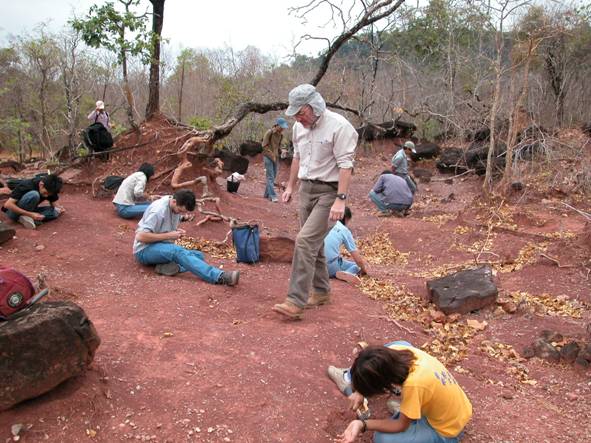
The team looking for fossils at Phu Phok (Thailand). Most specimens are quite small (less than a centimetre) and visible, scattered on the ground, when the sediment is washed out by the rain. Credit: Romain Amiot/Lyon University 1.
Each egg was scanned at very high resolution - each pixel measuring just 5 microns or the width of a strand of spider silk.
The analysis of the scanning results was a lengthy process. Using 3D software, each bone or fragment of bone, in each embryo, was analysed individually. Models of the bones were produced with a 3D printer and painstakingly pieced together to form a 3D reconstruction of the embryo.
Reconstruction of the anguimorph lizard embryonic skull using bones from the two best preserved eggs. Credit/ESRF. V. Fernandez.
Vincent Fernandez explains: "each time we had a new bone virtually extracted, we had a new clue about the nature of these embryos. We created a 3D print-out of each piece of the skeleton, and it took us several months of work before we could solve this scientific enigma.”
The eureka moment came during another unrelated experiment at the ESRF. Dr. Paul Tafforeau (head of the ID19 beamline, ESRF) recalls “a team came to study the inner ear of modern lizards and after their experiment they were looking at a 3D reconstruction of a common wall lizard. I immediately recognised one of the bones that we also had seen in the fossil embryos from Thailand: it was the quadrate, a bone for the articulation of the jaw.” After finding that cornerstone piece, all the elements of the skeleton fitted together like a jigsaw puzzle. "The embryos died at a stage when their skeletons were strong enough to fossilize. Exceptionally preserved, were recognized numerous diagnostic features that made it possible for us to disclose the issue about their true identity. They belong to the mighty group of the anguimorph lizards." says Dr.Martin Kundrat (Uppsala University/Czech Academy of Sciences).
The discovery of anguimorphs in hard-shelled eggs comes as a considerable surprise. Until now, only geckos were known to lay hard-shell eggs and most lizards lay soft-shelled eggs. The identification of the Thai embryos has an important scientific impact, providing essential information for our understanding of the evolution of reproduction in lizards and the diversity of eggs produced by different lineages of lizards.
The hard-shelled eggs of geckos were thought to be an evolutionary novelty within modern lizards. Consequently, all fossil eggshells resembling those of modern geckos were tentatively attributed to this group of wall-climbing lizards. This new discovery, however, shows that the evolution of lizard reproduction is more complicated than previously thought.
|
|
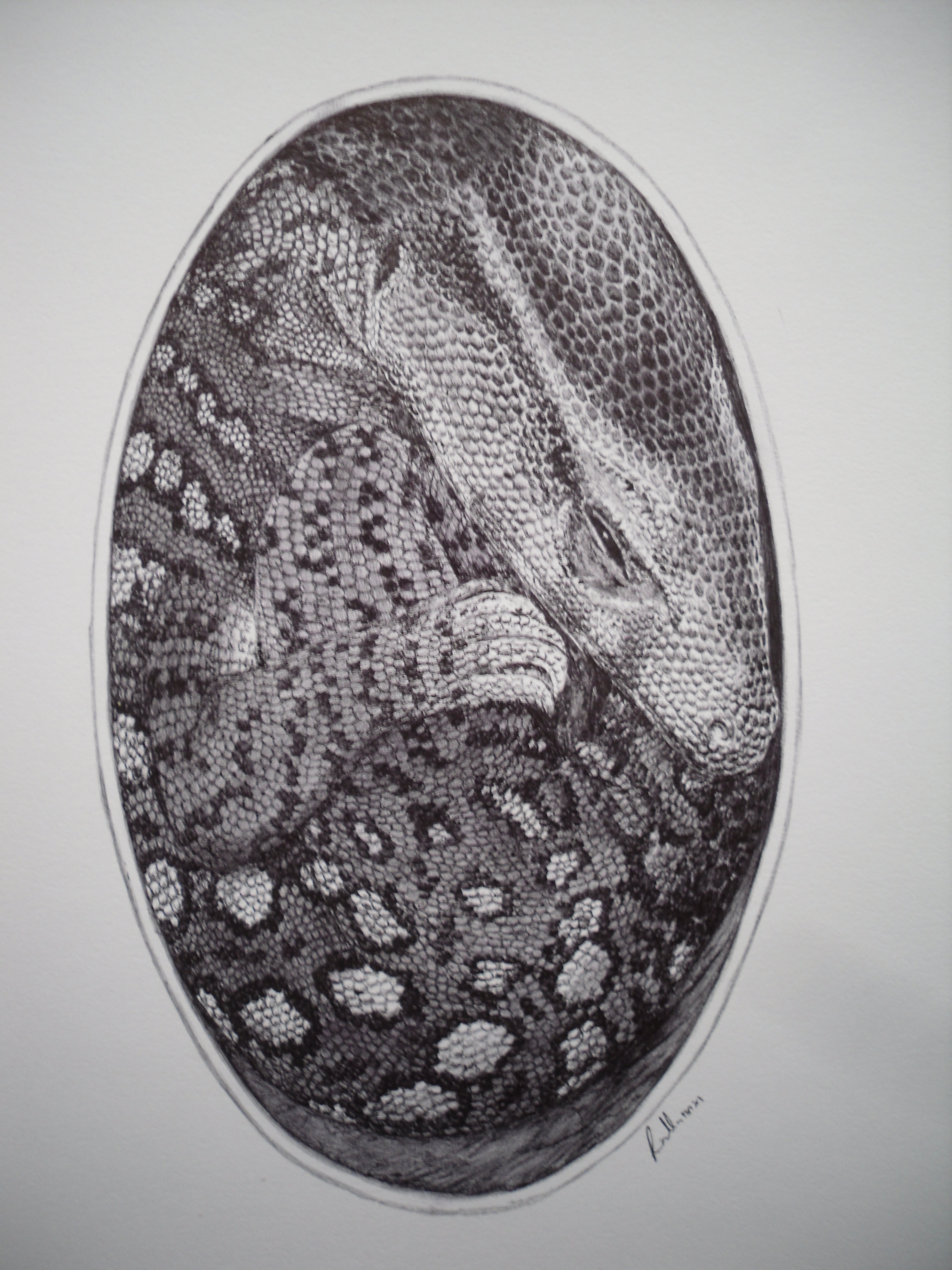
Artist's impression of the anguimorph lizard embryo in its egg. Full copyright is assigned to Vladimir Rimbala - the reconstruction was made under supervision of Martin Kundrat. |
Today's technological innovations enable scientists to see what was previously invisible
The eggs were discovered in 2005. So why has it taken so long to discover the true identity of their occupants?
Vincent Fernandez, who has led the research on these fossilised eggs since choosing them as a topic for his thesis in 2007, explains that although synchrotron X-ray techniques were available back then, algorithms and computers were a limit to be able to see the smaller bone fragments. The width of some bones inside these eggs only measure 10 microns: just two pixels across with a resolution of 5 microns. Vincent Fernandez recalls “In 2007, we had to scan the eggs at a resolution of 16 microns first. Not that we could not achieve higher resolution but even a compressed version of the data from a scan at 5 microns on these eggs was about 20 Gb, which was way too large for our best computers with 8 Gb of RAM. Thankfully the scans at 5 microns arrived early enough to include them in my PhD.” With this resolution, the egg could not be scanned as one piece but had to be examined in two fragments and the results "stitched" together to provide an image of the whole. Vincent Fernandez adds “This technique that allows us to double our field of view was non negligible for the whole computing process and it took quite some time to get the final data set ready.”
Finally, after tackling one obstacle after the other the data of these eggs could finally be analysed. Vincent Fernandez concludes “The 3D software we used to virtually extract the bones from the matrix had problems handling this data set. I was lucky enough that the next upgrade of the software was better adapted to this kind of work and that I could eventually discover the oldest lizard embryos in their eggs.”
Today the ESRF is one of the best facilities in the world for paleontological studies, not only for the variety of techniques that can be used to examine samples but also for the computing infrastructure and capacity required to handle the data generated. In 2008 at the ESRF, and still in many facilities around the world today computer ram is in the region of 64 Gb. Today, ID19 has two 512 Gb computers placing it at the very top of the scale for data collection and analysis. In 2012 with traditional techniques one egg was scanned and showed 62 bones. With the ESRF's better signal to noise ratio and using X-ray Phase Contrast technique, the same egg showed a clear outline of 265 bones.
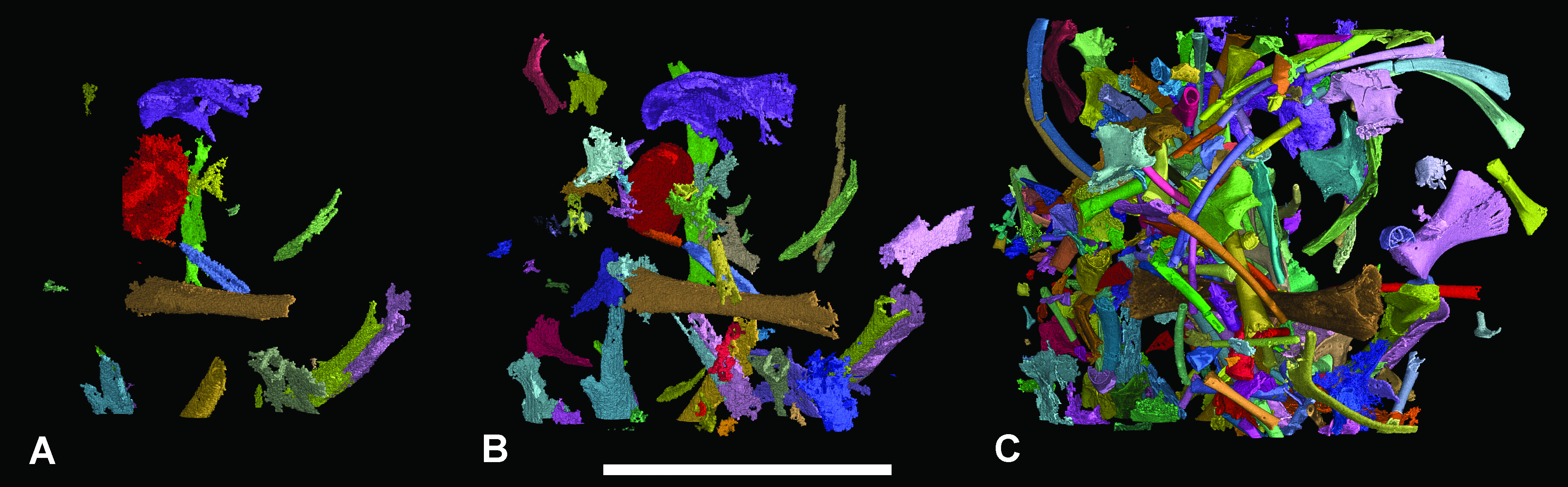
This figure shows a comparison of virtually extracted bones from one of the fossilised lizard eggs using different X-ray microtomographic techniques. A: with conventional microtomography (µCT) 21 bones are visible. B: with Absorption synchrotron microtomography (SR-µCT) 62 bones are visible. C: with propagation phase contrast X-ray synchrotron microtomography (PPC-SR-µCT) 265 bones are visible. Credit: V. Fernandez, microscopy and microanalysis 2012.
Download the press release (PDF)
Related articles:
Phase Contrast Synchrotron Microtomography: Improving Noninvasive Investigations of Fossil Embryos In Ovo. Microscopy and Microanalysis. Vincent Fernandez, Eric Buffetaut, Eric Maire, Jérôme Adrien, Varavudh Suteethorn and Paul Tafforeau (2012). View abstract
Top image: The tiny fossilised egg studied at the ESRF with colouring to show the embryonic bones preserved in the egg. Credit: E. Buffetaut/V. Fernandez
partners


European Synchrotron Radiation Facility - 71, avenue des Martyrs, CS 40220, 38043 Grenoble Cedex 9, France.